The “Laser Sources, Optical Radiation Characterization and Advanced Optical Imaging” cluster is a technical platform that brings together a range of cutting-edge photonics equipments, including laser chains, temporal, spectral and spatial metrology tools, and imaging tools, particularly for non-linear imaging. The services offered are based on commercial equipment or equipment developed in the laboratory as part of both fundamental projects (e.g., the study of nonlinear effects in optical guides) and applied projects (e.g., multiphoton imaging on living tissues).
2-photon fluorescence (2PEF) and second harmonic generation (SHG) microscopy
Olympus FV1200mpe (Coherent Chameleon Ultra laser source)
Multiphoton microscopy is an imaging technique that exploits the high power of short-pulse laser sources to generate multiphoton interactions in matter. The probability of occurrence of the phenomena and the longer wavelengths used (compared with linear imaging) enable intrinsic confocality and better tissue penetration (typ. 500 µm for 2-photon processes). What’s more, the high powers involved also make it possible to detect endogenous responses in biological samples, i.e. without the need for prior labeling.
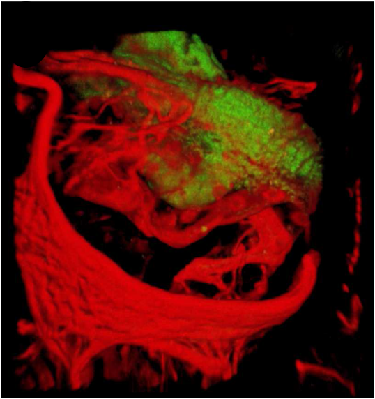
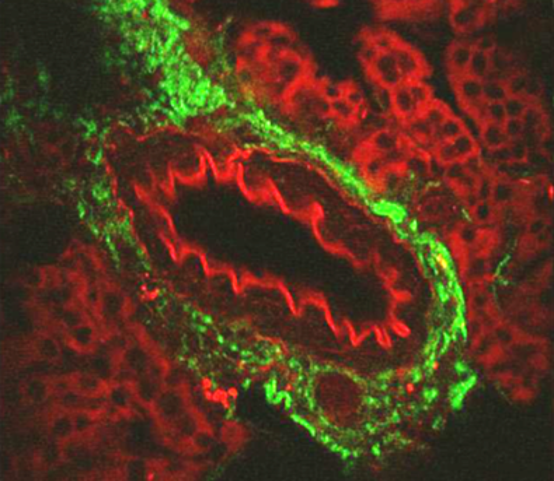
Examples of biological samples, 2-photon fluorescence in red (mainly elastin), second harmonic generation in green (collagen):
 |
 |
Lung artery |
Kidney artery |
Raman et CARS microspectroscopy
HORIBA LabRAM HR (633 nm source for Raman modality and ns 1064 pumped SC for CARS)
Raman spectroscopy is a technique used for determining the chemical composition of a sample. It involves illuminating the sample with a laser beam (usually visible and continuous) and detecting the scattered light. The spectrum of this scattered light reveals intensity peaks corresponding to the resonance of the sample’s molecular vibrations. The wavelength shift between the laser signal and that of the peak in question is then characteristic of a molecular bond present in the sample. In CARS (Coherent Anti-Stokes Spectroscopy), the sample is excited by two laser beams whose wavelength difference coherently excites the targeted molecular bond. This non-linear process provides a particularly high level of sensitivity. The technique used in the XLIM laboratory exploits the advantages of supercontinuum sources to combine two sources, one of which has a broad spectrum, to “read” a wide range of the vibrationnal spectrum of the sample without the need for wavelength scanning.
Microspectroscopy then involves coupling one of these techniques with spatial scanning in a microscope in order to reconstruct a “chemical” image of the sample.
To meet the scientific and technical challenges presented by an ever-increasing number of applications, laser sources must be able to offer a maximum of versatility in terms of wavelength, energy, repetition rate and pulse duration. The combination and synchronization of different types of radiation also opens the way to numerous fields of exploration in areas such as the characterization of non-linear phenomena in materials or the development of new imaging modalities.
Available sources:
Coherent Chameleon Ultra (760 to 1000 nm, 200 fs, 80 MHz) Amplitude Tangerine HP2 (1030 nm, 250 fs – 100 ps, 250 µJ, single shot to 40 MHz)
- 515 nm, 343 nm, 257 nm, 250 fs – 100 ps, 5 µJ, single shot to 40 MHz
- 200 à 2600 nm, 250 fs, single shot to 1 MHz
Spectral characterization
Optical spectrum analyzers :
- Anristu MS9710B (650 – 1750 nm)
- Yokogawa AQ6374 (350 – 1750 nm), AQ6375B (1200-2400 nm) et AQ6376 (1700 – 3400 nm)
Spectro-monochromator Newport + source Oriel (VIS to > 20 µm) Spectrograph ANDOR Kymera 193i (UV – NIR)
Temporal characterization
Fast oscilloscope Tektronix DPO77002SX ATI (70 GHz, 200 GS/s, 45 GHz photodiode ) Autocorrelator APE (20 fs – 5 ps, 700 – 1100 nm)
Spatial characterization
Analyseur de profil de faisceau (400 – 2100 nm) Caméra à détecteur InGaAs (900 – 1700 nm) Near field – far field characterization (400 – 1100 nm)
Spectro-temporal characterization
FROG Femtoeasy MS-FROG-SP (5 fs – 40 ps, 1500 – 2100 nm)
“Label-free discrimination of muscle proteins based on non-resonant background signature of multiplex-coherent anti-Stokes Raman scattering imaging”, M. Nafa et al., J Phys. D: Appl. Phys. 58 (2025)
https://doi.org/10.1088/1361-6463/ae1f9c
Boussafa, L. Sader, V. T. Hoang, B. P. Chaves, A. Bougaud, M. Fabert, A. Tonello, J. M. Dudley, M. Kues, B. Wetzel, “Deep learning prediction of noise-driven nonlinear instabilities in fibre optics”, Y, Nature Communications, 16, 7800 (2025)
https://doi.org/10.5281/zenodo.15179897
Tigran Mansuryan, Nicholas Bagley, Remy Boulesteix, Yago Arosa, Katarzyna Krupa, Benjamin Wetzel, Bruno P. Chaves, Stefan Wabnitz, Vincent Couderc, Alejandro Aceves, and Alessandro Tonello, “Nonlinear flying focus pulses for ultrafast 3D nonlinear microscopy,” Optica 12, 1192-1199 (2025)
https://doi.org/10.1364/OPTICA.561661
Zoltan S, Jonathan C, Jeremy M, et al. A novel histological occlusion classification for coiled aneurysms based on multiphoton microscopy. Interventional Neuroradiology. 2023;31(3):324-332
https://doi.org/10.1177/15910199231157926
Agenda Tous les événements