
Lymphoma: understanding the very early stages of its development
An innovative scientific article published in the prestigious journal Blood Advances sheds light on the fundamental mechanisms behind certain lymphomas and positions our laboratory at the forefront of research into cancers of the immune system.
New insights into the REL gene
The 2MB2C team at the CRIBL laboratory has highlighted the key role played by the REL gene in germinal centre B cells — the antibody “factories” located in our lymph nodes. They demonstrated that genetic deregulation of the REL gene in germinal centre B cells creates a lasting reservoir of lymphoma precursor cells, particularly in the diffuse large B-cell lymphoma subtype of germinal centre origin (GCB-DLBCL). Using an original ‘dual-colour’ model, the authors were able to track, for the first time, the competitive evolution of B cells overexpressing REL compared to healthy cells during chronic antigenic stimulation.
A competitive and persistent mechanism
The study shows that REL overexpression promotes B cell expansion, class recombination and plasma cell differentiation. More strikingly, this alteration confers a lasting competitive advantage, allowing these cells to persist, recirculate and even expand clonally within the body.
Towards a better understanding of lymphomagenesis
The results demonstrate that REL deregulation acts as a fundamental oncogenic event in the early stages of B-cell transformation and lays the foundation for a long-term reservoir of pre-lymphomatous cells. These findings open up new avenues for early therapeutic targeting and prevention of lymphoma development in at-risk populations.
Laboratory outreach
This publication reinforces the laboratory’s international recognition and highlights the excellence of its team in understanding the early stages of lymphoid cancer. It illustrates the fundamental impact of translational research on diagnostic and therapeutic innovation in haematology.
This work was supported by the ARC Foundation for Cancer Research and the League Against Cancer.
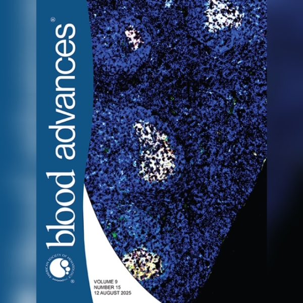
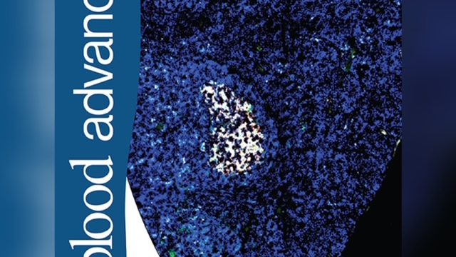
Cover image
The icing on the cake was that one of the immunofluorescence images from the article was selected to illustrate the cover of the August 2025 issue of Blood Advances, volume 9, no. 15.
